22 Dec, 2025
22 Dec, 2025

This article is medically reviewed by Dr. Tushar Kumar Mohapatra, Senior Consultant – Nuclear Medicine, HCG Panda Cancer Hospital, Cuttack.
PET-CT (Positron Emission Tomography/Computed Tomography) is a nuclear medicine imaging technique that combines the structural information from the CT scan with the metabolic or biochemical activity information from the PET scan. It is a cancer diagnosis test.
A software system combines PET and CT imaging data for two—or three-dimensional image reconstruction. These combined scans help oncologists pinpoint abnormal metabolic activity and devise the best treatment plan for various cancers. Also, PET-CT may provide a more accurate diagnosis than the diagnosis obtained from PET or CT alone.
PET-CT imaging helps in staging, treatment planning, treatment monitoring, restaging, and recurrence evaluation. One of the common questions cancer patients ask is, “Can cancer be detected in a CT scan?”
It is important to understand the advantages and limitations of CT scans and how these limitations are overcome by using CT scans with PET scans.
Following are some of the points of difference between the positron emission tomography scan and the CT scan:
A PET scan for cancer screening is a useful method. This nuclear medicine imaging method involves injecting a radioactive tracer into the patient's bloodstream.
CT scan, on the other hand, uses X-rays to create detailed images of the body's internal structures.
"PET-CT scan dual approach significantly enhances our ability to detect, stage, and monitor diseases,
particularly cancer. It allows for precise treatment planning and early intervention, which are crucial
for successful patient outcomes."
Dr. Tushar Kumar Mohapatra
Sr. Consultant – Nuclear Medicine
HCG Panda Cancer Hospital
During PET scans for cancer, the underlying mechanism detects tissue metabolic activity.
On the other hand, the CT scan focuses on creating images of various body organs. The absorption of X-rays by the tissues depends upon their density.
Watch this video where we discuss how PET-CT works:
The images obtained in the PET test for cancer are color-coded to depict the areas of high and low tracer concentration and high and low metabolic activity. The cancer cells have a higher metabolic rate than normal cells, thus appearing as bright spots on PET images.
The images obtained with the CT scan are highly detailed and show the body's anatomical structures. These images are useful in identifying abnormalities in the structure of tissues and organs.
PET scan uses include disease diagnosis and treatment evaluation in the fields of oncology, neurology, and cardiology. They help detect and stage tumors, study brain function, and evaluate myocardial perfusion.
A CT scan spots internal injuries, detects tumors, guides biopsies, assesses heart disease, evaluates stroke and brain injuries, and identifies bone fractures.
The advantages of PET scans for cancer include providing functional and metabolic information crucial for detecting cancer, being highly sensitive, and being useful in diagnosing neurological and cardiological conditions.
The advantages of CT scans include quick and wide availability, non-invasiveness, and relatively lower risk. They also provide detailed images of the blood vessels, bones, and soft tissues.
The disadvantages of PET scan tests are exposure to radioactive tracers, relatively high cost, limited availability, and lower spatial resolution compared to CT scans.
The disadvantages of CT scans are exposure to ionizing radiation and the limited ability to differentiate between tissue types.
| Characteristic | PET Scan | CT Scan |
|---|---|---|
| Basic Characteristics | Nuclear medicine imaging involving radioactive material | Images are taken through X-rays |
| Focus | To determine the metabolic activity of the tissues | To determine anatomic abnormalities |
| Clinical Applications | Used in diagnosing cancer, studying brain function, and evaluating myocardial perfusion. | Used in diagnosing internal injuries and bleeding, detects tumors, guides biopsies, and identifies bone fractures |
| Advantages | Provides functional and metabolic information crucial for detecting cancer | Quick and wide availability, non-invasiveness, and lower risk |
| Disadvantages | Exposure to radioactive tracers, relatively high cost, and limited availability. | Exposure to ionizing radiation and the limited ability to differentiate between tissue type |
A PET-CT scan helps both patients and doctors in numerous ways:
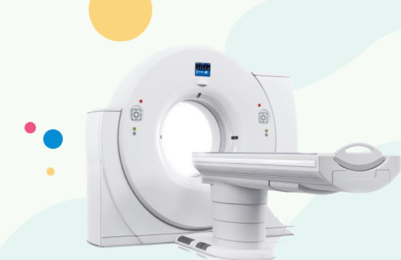
The PET-CT scan machine combines the technologies of PET and CT scans. It is a cylindrical machine with a tunnel at the center. The device looks similar to a CT scanner but has the additional components of a PET scanner.
The central part of the machine is known as the gantry. The gantry comprises an examination table that moves in and out of it. The examination table has straps to fix the patient in one position during imaging.
The PET-CT scan machine comprises the CT, PET, and central console. Patients see the gantry as a large circular hole into which they will slide on the examination table. The machine also has a technician interface with multiple monitors displaying real-time images and diagnostic information.
The integration of PET and CT scan technologies allows for simultaneously obtaining anatomical and metabolic data. It also ensures patients' comfort, as they do not need to be moved from one machine to another.
The doctor recommends the PET-CT scan for those suspected of having metastatic tumors.
In the beginning, the patient is provided with advance instructions related to eating and drinking. The doctor also asks the patient about their medical history, including allergies and current medications.
"Proper preparation for a PET-CT scan is crucial for accurate results. Patients should follow all instructions regarding fasting and medication. Informing us about allergies and medical history helps ensure safety and effectiveness. Wearing comfortable, metal-free clothing and staying hydrated are also important. These steps maximize image clarity and contribute to a smooth, successful procedure.”
Dr. Tushar Kumar Mohapatra
Sr. Consultant – Nuclear Medicine
HCG Panda Cancer Hospital
Upon arrival at the hospital, the patient is asked to change into hospital gowns. The doctor or radiologist injects a small amount of radioactive substance into the patient's vein.
After the injection, the patient is advised to wait for about 30-60 minutes for the radioactive tracer substance to distribute itself throughout the body.
The patient is asked to lie on the examination table and is recommended to stay as still as possible to obtain clear images. The scan usually begins with a CT scan. After the CT scan, the patient undergoes a PET scan. The images obtained with the PET and CT scans are combined using computer software.
Patients should follow the radiologist's instructions for better-quality imaging during a PET-CT test. Some of these instructions are:
PET-CT scans have some limitations. The method involves exposure to ionizing radiation. Repeated scans may lead to cumulative radiation dose, which is a concern, especially in young patients and patients requiring multiple follow-up scans.
The availability of PET-CT scans is also a challenge. As the equipment is expensive, many healthcare facilities in Tier II and Tier III cities may not have it.
A PET-CT scan for cancer is not a confirmatory diagnostic test; a biopsy is usually required for a confirmatory diagnosis.
It is not suitable for all the patients. PET-CT scans are not done in pregnant and breastfeeding women or patients with severe claustrophobia.
HCG Panda Cancer Hospital, a leading cancer hospital in Cuttack, has an advanced diagnostic facility for a comprehensive examination of patients suspected of various cancer types. The PET-CT scan for cancer is available at the center to diagnose various types of cancer. The radiologists handling the machine are highly trained to optimize the technique and improve the quality of the results.
A PET-CT scan for cancer combines positron emission tomography (PET) with computed tomography (CT) to offer detailed images of metabolic activity and anatomical structures, optimizing cancer detection, staging, and treatment planning.
This technique provides precise information about abnormalities, aiding in early diagnosis and effective treatment plans. Despite its benefits, it involves radiation exposure and may not be suitable for everyone, such as pregnant women. A full-body PET scan for cancer is usually recommended to determine the extent of disease spread.
Author Bio : Dr. Tushar Kumar Mohapatra
MBBS, MD (Nuclear Medicine) Sr. Consultant – Nuclear Medicine
Dr. Tushar Kumar Mohapatra is an experienced nuclear medicine specialist at HCG Panda Cancer Hospital, a leading cancer hospital in Odisha. He specializes in diagnostics, therapeutic intervention, radiation safety, and tele-radiologic reporting. He has a special interest in PET CT studies, PET-based radiotherapy planning, iodine therapy, and more. He is also experienced in performing procedures like SISCOS and SISCOM on epilepsy patients. He is a highly respected professional in the field and is known for his expertise in the field of nuclear medicine.
Feel free to reach out to us.