23 Dec, 2025
Feel free to reach out to us.
23 Dec, 2025

This article is medically reviewed by Dr. Sandeep Kumar, Consultant - Surgical Oncology, HCG-Abdur Razzaque Ansari Cancer Hospital, Ranchi.
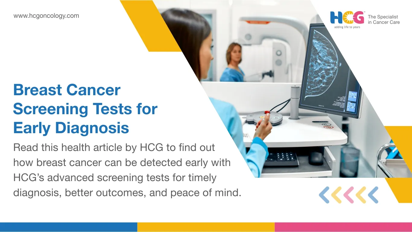
Breast cancer screening involves a group of medical tests used to detect breast cancer at an early stage, generally before the symptoms appear.
The primary aim of breast cancer screening is early diagnosis and to identify the cancer when it is localized, small, and easy to treat with a high degree of favorable prognosis.
Breast cancer detection at an early stage significantly enhances the outcomes and survival rates while lowering the requirement for aggressive therapies.
Common breast cancer screening methods are mammography, which uses low-dose X-rays to detect tumors or abnormalities; breast magnetic resonance imaging for detailed images for high-risk women; and molecular breast imaging, which is an advanced technique to highlight the cancerous cells.
Apart from the above methods, clinical breast examinations performed by doctors and self-breast examinations also help detect breast changes at early stages.
Breast cancer screening recommendations depend on the risk level, age, and family history. For most women, a regular mammogram is recommended at age 40. However, those at higher risk should start the screening early.
Regular and recommended screening for breast cancer significantly lowers mortality by ensuring early intervention.
The breast cancer survival rate is significantly increased because of early detection through screening. This section is focused on answering the question, "How to test for breast cancer?"
Screening tests for breast cancer include various imaging and non-imaging techniques to detect cancer even before the symptoms appear.
A mammogram is the most commonly used and widely recommended for screening breast cancer.
It involves low-dose X-rays to find abnormal growths or microcalcifications. These characteristics may suggest cancer even before the actual cancer symptoms appear.
Mammograms are effective in the early diagnosis of breast cancer.
Regular mammograms in women aged 40 and above, especially those at elevated risk for breast cancer, significantly lower the deaths due to breast cancer.
At HCG, different types of mammography, such as digital mammography, CR mammography, 3D mammography, etc., are recommended for accurate detection of breast cancer.
When the doctor requires detailed images of the breast tissue, the patient is advised to undergo a breast MRI. The procedure includes the magnetic fields and the contrast dye.
It is particularly useful in women with dense breasts or with a strong family history of breast cancer.
Breast MRIs can detect abnormal growth that mammograms miss. However, it is generally not used alone due to its cost and higher false-positive rate.
MBI is an advanced nuclear medicine procedure that involves a small amount of radioactive tracer to detect cancerous cells.
It complements the mammography and detects cancer cells in dense breast tissue.
It improves overall testing sensitivity and allows oncologists to obtain an accurate diagnosis of hidden or small tumors.
Routine clinical breast examinations performed by the healthcare provider and regular breast self-examinations help women to become familiar with the appearance of their breasts. Thus, any changes in their breast are immediately identified and encourage medical consultation.
Women should not ignore any changes, unusual symptoms, or lumps in their breasts. They should seek advice on undergoing diagnostic measures for breast cancer.
Breast cancer screening guidelines focus on detecting breast cancer in the early stage, when the treatment is most effective, with a higher survival rate. The recommendations slightly vary based on age, risk factors, and health history.
1. For women at average risk of having breast cancer, women between 40 and 44 years of age should undergo an annual mammogram. Women between the ages of 45 and 54 should undergo an annual mammogram. Screening is recommended every two years after age 55.
2. Healthcare providers may further schedule the screening duration based on the overall health condition.
3. For women at a higher risk of developing breast cancer, e.g., those with a family history of cancer, BRCA1 or BRCA2 gene mutations, or a history of chest radiation, the annual mammograms and breast MRI should start at 30 years of age or as advised by the physician.
4. In addition to the recommendations for undergoing imaging scans, clinical breast examinations and self-examination of the breasts are recommended at all ages.
5. Women should immediately report any changes in the appearance and texture, presence of lumps, and nipple discharge to their gynecologists.
6. Regular screening, along with a healthy lifestyle and awareness about breast cancer symptoms, significantly improves the chances of detecting breast cancer at early stages and obtaining better treatment outcomes.
7. Intervals between two screenings should be personalized after a medical consultation.
Advantages of breast cancer tests for screening are:
Screening for breast cancer assists in the identification of tumors at an early stage, generally before the symptoms appear. It allows for timely and more effective treatment. Detecting cancer early enhances survival rates and treatment success.
Cancer diagnosed at early stages requires less extensive surgery, reduced chemotherapy doses, and fewer radiation therapy sessions. It results in faster recovery and fewer side effects.
Routine screening, especially through mammography, lowers the breast cancer-related deaths, as it detects the cancer at an early stage when it is most curable.
Screening helps in routine evaluation, close monitoring, and early management for a woman with a family history of breast cancer or genetic predisposition, thereby lowering the long-term risks.
Routine screening provides reassurance to women with normal results and motivates them to take proactive measures for health management. It improves awareness and boosts confidence in breast health.
Some of the risk factors that make the test for breast cancer screening essential are:
Age is directly associated with the risk of breast cancer. The breast cancer risk increases with age, especially in women over 40 years of age.
Regular screening is important, as early detection of breast cancer significantly improves the overall outcomes in older women.
Women with a known BRCA1/BRCA2 gene mutation or who have had their close relatives diagnosed with breast cancer are at higher risk.
Screening in this population should start early, and they should undergo both mammography and breast MRI.
Periodic screening is vital in those women who have early menstruation (before 12 years of age), late menopause (after 55 years of age), or are on long-term hormone replacement therapy, as these are at elevated breast cancer risk.
Lifestyle factors, such as obesity, sedentary habits, and alcohol consumption, elevate the risk of breast cancer.
Screening in women with these risk factors helps in detecting the abnormalities at an early stage.
Women who have received chest radiation or have a history of benign breast disease are at elevated risk of breast cancer and should undergo regular screening to monitor for malignancy.
Breast density refers to the volume of fibroglandular tissues compared to the fatty tissues present in the breast. Women with dense breasts have a higher volume of fibroglandular tissue compared to fat tissue.
The fibroglandular tissues project themselves as a white mass in the mammogram, similar to the tumors. Thus, it is hard to detect the abnormalities in women with dense breasts, resulting in reduced mammogram accuracy. It enhances the likelihood of false positive results or missed cancers.
Women with dense breasts are also at an elevated risk of breast cancer. Therefore, the doctors usually recommend additional imaging tests, such as MRI, MBI, or ultrasound, for more accurate screening in this population setting.
A woman may expect the following while undergoing a breast cancer screening test:
During a mammogram, the woman is asked to stand in front of an X-ray machine. The radiologist gently compresses each breast between two plates.
The pressure may last only for a few seconds and allows the radiologist to take clear images of the breast tissue. You may experience mild discomfort; however, the procedure is quick and safe.
If your oncologist advises you to undergo a breast MRI, you are instructed to lie inside a scanner. You may have a contrast dye to highlight the breast tissues. The procedure is usually painless and takes about 30 to 45 minutes.
During a clinical breast examination, a healthcare provider physically examines the breasts for detecting any lump or abnormality.
You can resume your normal activities immediately after the tests. Results are generally shared with you within a few days.
If any adverse results are found, the doctor may advise you to undergo further imaging tests or a biopsy to confirm the disease. Regular screening results in early and accurate detection.
Accurate interpretation of the screening results of the breast tissues is important to avoid missing any abnormality. After the screening tests are done, the experienced radiologist evaluates the scanned images for any signs of abnormal masses, tissue, or calcifications.
The results are categorized using the BI-RADS (Breast Imaging Reporting and Data System), which ranges from 0 to 6.
The zero value suggests that additional tests are needed, while 1 or 2 indicates normal or benign findings.
A score of 3 suggests a probably benign growth and may require a short-term follow-up.
A score of 4 or 5 indicates suspicion or high likelihood of cancer, and a score of 6 confirms a known malignancy.
Based on the above scores, the oncologists examine the findings in detail and advise the next steps. The next steps may include routine monitoring, repeat imaging, advanced imaging, or a biopsy.
The possible risks or limitations of screening for breast cancer include:
In some cases, the screening tests may detect abnormalities that are not cancerous.
These false positive tests may lead to unnecessary imaging and biopsy. It also causes anxiety and emotional distress among individuals.
Screening may also detect the slow-growing cancers that may not have the potential to spread.
Treating these tumors immediately may expose the women to unnecessary surgery, chemotherapy, or radiation therapy.
Mammogram uses low-dose radiation. Although the risk from this low-dose radiation is minimal, repeated exposure over time may slightly increase the overall risk of cancer.
Advanced screening tests, such as molecular imaging or breast MRI, are expensive and sometimes unnecessary because of the false positive results of mammograms. Further, they are not widely available and have limited accessibility for some women.
Despite these limitations, routine screening for breast cancer is important, especially for those at high risk of developing breast cancer.
Genetic tests are important in personalized breast cancer screening. The oncologists may recommend genetic testing to assess the risk of breast cancer in certain women based on their eligibility. Genetic testing has several advantages.
Genetic testing detects mutations in genes, such as BRCA1, BRCA2, PALB2, and TP53.
Mutations in these genes increase the risk of breast cancer. Women with these mutations are closely monitored and are under long-term surveillance.
Based on the genetic test results, the oncologists customize the screening schedules, such as recommending annual mammograms and MRIs at a younger age.
It ensures early detection of breast cancer followed by timely treatment.
Genetic testing results allow oncologists to guide the preventive steps that may include lifestyle changes, preventive surgeries, and risk-reducing medications in those with a high risk of developing breast cancer.
Once a woman is detected with a high risk of having breast cancer through genetic testing, the other family members are also advised to undergo genetic testing. It will allow them to take preventive or screening measures.
Decision-making is unusually precise when the data is in hand. Once the genetic risk status is identified, the women are able to make informed choices about their health, thereby improving the long-term outcomes.
In India, the cost of screening for breast cancer varies significantly.
It depends on the type of test and the facility location. In private hospitals, a screening mammogram generally costs between ₹1,500 and ₹3,000, while a digital or 3D mammography costs slightly more and is between ₹3,000 and ₹6,000.
Breast MRI or molecular breast imaging is more advanced, and the cost of them is usually between ₹8,000 and ₹15,000. In government hospitals, the screening tests are either free or available at subsidized rates.
Several insurance providers allow the coverage of mammograms only if advised by the doctor. However, they may not cover the routine preventive screening.
Some wellness or preventive health plans specially designed for women's health may include the annual breast screening benefits.
Technology is a continuously evolving domain. Several advancements have occurred in the field of breast cancer screening. These technologies improve the accuracy and accessibility of breast cancer screening.
It is an advanced mammography. It captures multiple slices of the tissue, reduces tissue overlapping, and improves detection, especially in those with dense breasts.
Studies suggest that 3D mammography has a higher cancer detection rate and reduced recall rate.
An AI system assists the radiologists by analyzing the mammograms and other imaging to detect abnormalities at a higher speed and accuracy.
A large study suggests higher detection when AI assists the radiologists.
Non-invasive screening tools, such as thermal imaging, detect the heat and vascular changes in the breast tissue. The technique is especially useful in areas where access to standard mammography is limited or when the patient is apprehensive about the radiation exposure.
Advanced techniques, such as contrast-enhanced spectral mammography, allow radiologists to better visualize the vascular changes and hidden lesions, especially in those with higher risk or dense breasts.
Advanced technologies in breast cancer screening offer greater precision, early detection, and accessibility, thereby improving the overall care in breast cancer.
Together, these technologies are shifting screening toward greater precision, early detection, and accessibility, offering hope for improved outcomes.
Individuals choosing HCG Cancer Centre as their preferred center for comprehensive breast cancer care services have several advantages.
HCG Cancer Centre is equipped with state-of-the-art diagnostic tools, such as digital mammography, breast MRI, and molecular breast imaging. These technologies improve the detection accuracy, particularly in women with dense breast tissue.
Screening test of every patient is interpreted by extensively experienced oncologists and trained radiologists. This ensures accurate diagnosis and customized recommendations for further care.
The Preventive Oncology Centre at the HCG customizes the screening plans based on the risk profile, family history, and genetics, thereby improving early detection.
HCG Cancer Centre provides emotional support, patient-centric counseling, and a quick report to the individuals. It makes the process, from screening to diagnosis, smoother for the patients.
Screening is an important component of a comprehensive breast care strategy. It allows for early detection of breast cancer and improves treatment effectiveness and survival.
It also reduces the overall cost of treatment, as early-stage treatment is significantly less expensive than advanced-stage treatment. Breast cancer screening is recommended based on age, risk factors, and genetic predisposition.
Most women do not experience any side effects with screening. You should not ignore the symptoms of breast cancer.
You should book an appointment with your gynecologist and undergo tests for breast cancer detection.
Dr. Sandeep Kumar
Consultant - Surgical Oncology
MBBS, MS (General Surgery), MCh Surgical Oncology
Dr. Sandeep Kumar is a highly experienced and accomplished surgical oncologist, and he can be consulted at HCG - Abdur Razzaque Ansari Cancer Hospital, a leading cancer hospital in Ranchi. He specializes in the management of a wide range of cancers, including hepatopancreaticobiliary, gallbladder, breast and gynecological, thoracic, stomach, colorectal, soft tissue, and head and neck cancers. He is recognized for his technical excellence and compassionate patient care. His treatment approach is to provide a comprehensive, multidisciplinary approach, with an emphasis on the best possible outcome for each patient.
Appointment Link: Book an Appointment with Dr. Sandeep Kumar.