07 Nov, 2025
Feel free to reach out to us.
07 Nov, 2025

This article is medically reviewed by Dr. Sachin Gawde, Consultant - Nuclear Medicine, HCG Cancer Centre, Borivali.
Rapid and accurate diagnosis is the key to countering cancer, which is possible with imaging technologies.
In oncology, the three most common imaging techniques are PET, CT, and MRI, and each has its own strengths and limitations.
Being aware of the difference between CT scans, MRIs, and PET scans can help patients and their families make informed health decisions.
This blog article will focus on PET scanning, how it works, and how it compares with other scans, such as MRIs and CT scans, concluding with why it is hailed as an effective cancer-detection tool.
Positron emission tomography (PET) scan is a nuclear medicine imaging technique that helps specialists visualize the metabolic activity or functioning of tissues present in various parts of the body.
During the scan, a small quantity of radioactive material (tracer) is injected into the body. A tracer is a radioactive isotope tagged with a target molecule, which helps in assessing the active disease because cancer cells grow or multiply more rapidly than normal cells.
The PET scanner detects the radiation emitted from the tracer absorbed by cancer cells and constructs precise images to show how tissues or organs are functioning.
The PET/CT scan is the amalgamation of functional imaging details obtained from PET scanning and structural/anatomic imaging details from CT scanning.
The ultimate view obtained not only delineates abnormal activities but also exact locations.
This combination scan enhances diagnostic accuracy, especially in cancer; therefore, it has become a crucial tool in oncology.
Additional Reading: PET/CT Scan for Cancer Diagnosis
The procedure starts with the injection of a radioactive tracer into the patient’s vein.
After waiting for about 45-60 minutes for the tracer to circulate, the patient lies on a scanning table that moves through the PET/CT scanner.
The scan itself typically takes 30-45 minutes. The patient is required to remain still during the scan to ensure clear images.
The PET/CT scan is primarily used for:
A PET/MRI scan integrates the functional imaging capabilities of PET and the detailed soft tissue contrast of MRI.
This new technology gives very high-resolution imaging and is especially suitable for images of the brain, pelvis, and musculoskeletal system.
Nonetheless, the PET/MRI scanner is not as commonly installed as the PET/CT for the applications of special cases.
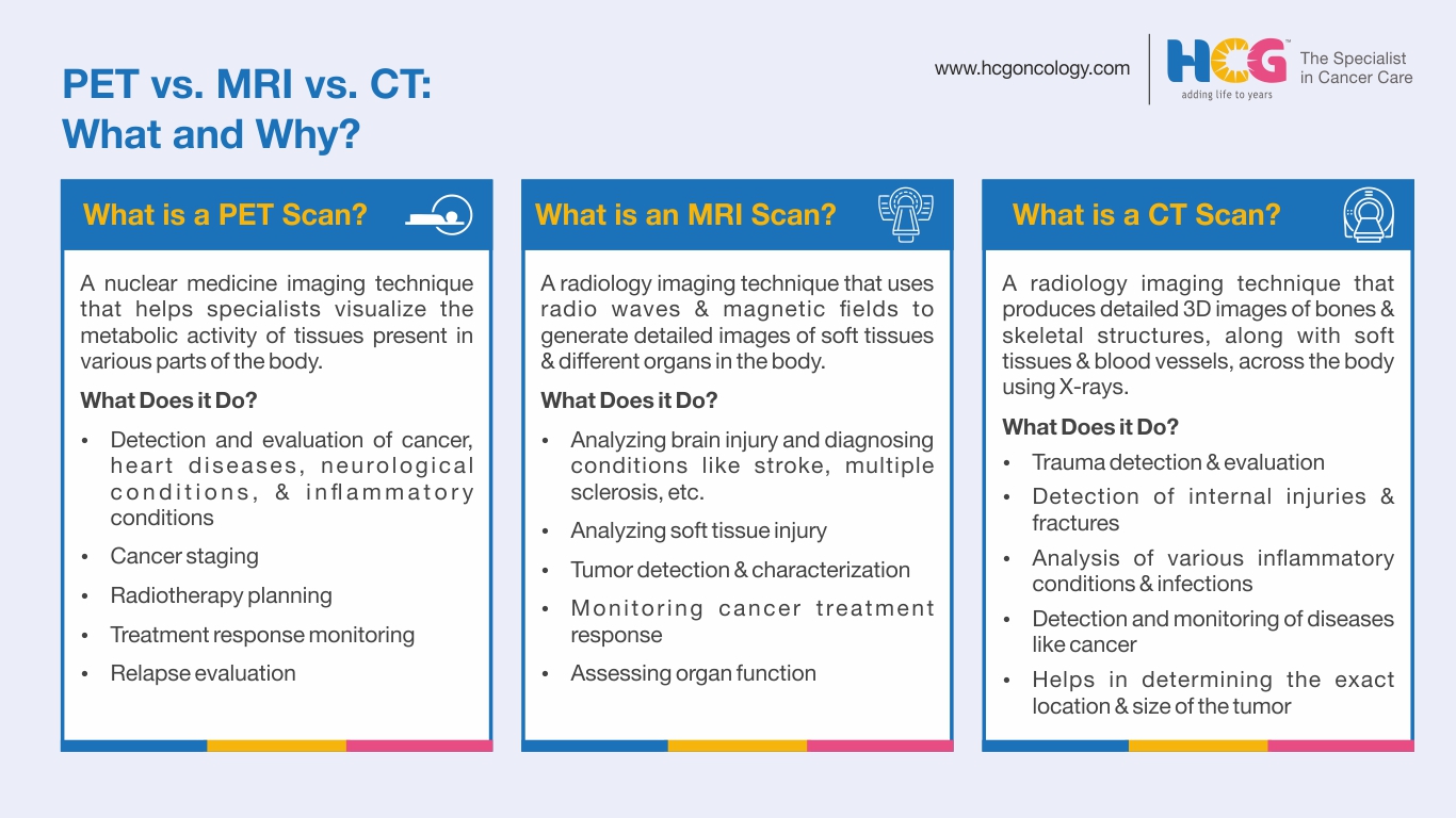
When evaluating PET scans vs. CT scans vs. MRI, it's important to understand their unique capabilities:
| Parameters | PET Scan | CT Scan | MRI Scan |
|---|---|---|---|
| Definition | Uses radioactive isotopes to visualize the metabolic activity of various tissues in the body. | Uses X-rays to create 3D images of different parts of the body. | Uses radio waves and magnetic fields to generate detailed images of soft tissues in different parts of the body. |
| What Do They Show | Cellular activities in various body parts and abnormal metabolic activity in case of any illnesses. | Bone and skeletal structures, along with soft tissues and blood vessels. | Detailed structures of soft tissues and organs, including muscles, the brain, and the musculoskeletal system. |
| Applications | For the detection and evaluation of abnormal metabolic activities indicating the presence of cancer, heart diseases, neurological conditions, and inflammatory conditions | For the detection and evaluation of trauma, fractures, infections, and certain tumor growths. | For the analysis of brain and spinal cord injuries, soft tissue damage, and tumors present in different parts of the body. |
| Radiation Exposure | Brief radiation exposure to the body cells from the radiotracer. | Moderate radiation exposure to the structures being scanned. | No radiation exposure. |
| Scan Duration | 60-90 minutes | 15-30 minutes | 15-90 minutes |
The above table addresses various queries like “the difference between an MRI and a CT scan,” “MRI versus PET scan,” and “the difference between CT and PET scans” in the best way possible.
Usually, before the scan, patients are asked to abstain from eating or drinking, except for water, for about 6-8 hours prior.
Blood sugar is monitored, especially in diabetes cases, to ensure accuracy during the scan procedure. Inform your physician regarding any medications or underlying health conditions.
A radiotracer will be injected into your body, and you will be asked to stay still for about 45-60 minutes to allow the radiotracer to circulate throughout your body.
Thereafter, the scanning procedure will start. You will be positioned on the patient couch, which will slide through the machine.
It is a pain-free procedure, but some people may feel slight discomfort as they are expected to lie still.
You can usually resume normal activities right after the scan. It is advisable to drink plenty of fluids to help flush out the tracer from the body.
The images will be interpreted by a nuclear medicine physician, and the findings will be made available to your doctor.
Additional Reading: What Should Patients Know About Their PET/CT Scan?
Generally, PET/CT scans are safe; however, this diagnostic method requires the use of radioactive tracers and X-ray radiation, which means exposure to small amounts of radiation.
Rare but possible allergic reactions to the tracer can also occur. Pregnant and breastfeeding women should inform their doctor beforehand to assess the risks.
Many assume that PET/CT is only used in the diagnosis of cancer. The role of PET/CT in managing cancers is only understood by a few.
Apart from cancer diagnosis, PET/CT imaging helps in cancer staging, treatment planning, and treatment response monitoring.
PET/CT enhances the precision in diagnosis, treatment, and response assessment for different types of cancer.
As a rule, a PET scan is acquired as whole-body imaging because cancer can spread from its site of origin to any other part of the body. This makes the PET/CT scan an important imaging tool in diseases like cancer.
HCG is a leading provider of comprehensive cancer care in India. Choosing HCG for your PET/CT scan ensures:
Understanding the difference between a PET scan and a CT scan for cancer, as well as an MRI vs. a PET scan for cancer, can guide better decision-making. When it comes to PET scans vs. CT scans vs. MRIs, each has its place in the diagnostic process.
The PET/CT scan stands out for its ability to merge function and structure/anatomy, offering unparalleled insights into the behavior of cancer in the body.
Still wondering, "Is a PET scan better than an MRI?" The answer depends on the specific clinical scenario. But for detecting cancer and assessing its spread, PET/CT scans often provide the most comprehensive picture.
Whether you're evaluating the difference between a CT scan, an MRI, and a PET scan or comparing an MRI versus a PET scan, it's clear that each tool is critical in its regard. HCG’s expertise and advanced facilities make them a top choice for anyone seeking clarity and confidence in their cancer journey.
Dr. Sachin Gawde
Consultant - Nuclear Medicine
MBBS, DRM (Diploma in Radiation Medicine), FANMB
Dr. Sachin Gawde is a nuclear medicine and PET/CT specialist with over 14 years of experience. He can be consulted at HCG Cancer Centre, one of the leading hospitals for cancer treatment in Borivali. His expertise lies in PET/CT imaging and administering high-dose radioisotope therapies for various conditions. He has reported on over 20,000 PET/CT scans and diagnostic nuclear medicine investigations. He takes time to understand each patient’s concerns and is known for creating a comfortable care setting for his patients.
Appointment Link: Book an Appointment with Dr. Sachin Gawde.