10 Apr, 2026
Feel free to reach out to us.
10 Apr, 2026
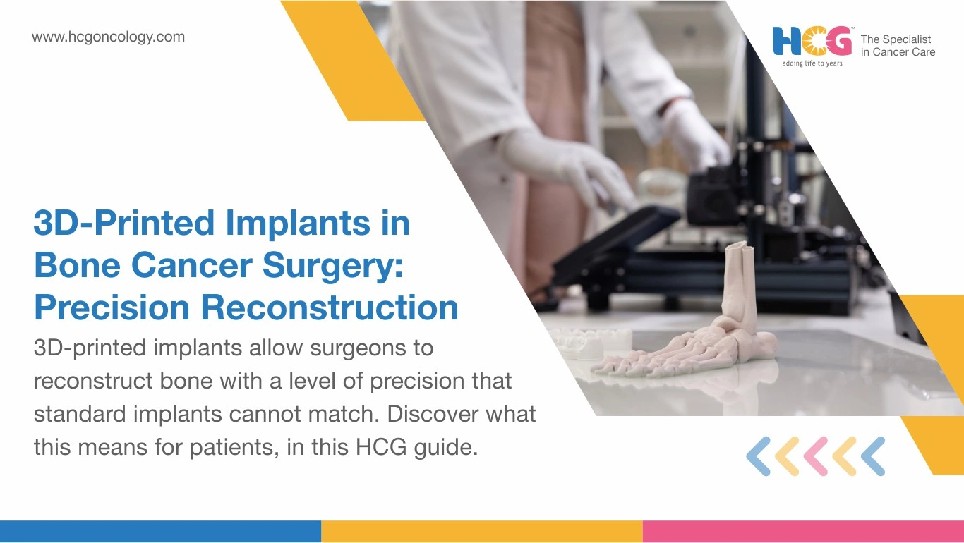
Losing a limb to cancer is a fear that sits quietly behind every bone tumor diagnosis. Patients do not always say it out loud, but it is there. What surgeons can offer today, though, is genuinely different from even a decade ago, and 3D printed implants in oncology are a large part of why.
The idea is straightforward. Every patient's bone structure is unique. So instead of working with a standard implant that has to be trimmed and adjusted in the operation theater, surgeons now scan the affected area beforehand and build a titanium implant to match it exactly. It arrives ready. It fits the first time.
For patients dealing with bone cancer, that precision matters, not just for the surgery itself but for recovery, for movement, and for life after treatment.
| Feature | 3D-Printed Custom Implant | Standard Prosthetic |
|---|---|---|
| Anatomical fit | Exact skeletal match | Approximate size range |
| Intraoperative adjustment needed | Minimal | Frequent |
| Biomechanical load pattern | Mirrors native bones | Can concentrate stress at junctions |
| Performance at complex sites | Strong (pelvis, proximal femur) | Limited in irregular anatomy |
Before the patient enters the operation theater, a significant amount of work has already happened.
It starts with imaging. A CT scan and MRI together produce a detailed, three-dimensional picture of the affected bone: its shape, its size, and exactly how much of it the tumor has involved. Engineers and surgeons use that data to design an implant that matches the patient's anatomy precisely, not approximately.
The implant is then printed in titanium, built up layer by layer using a process called selective laser sintering. What makes the implant more than just a metal piece is what is built into it. The implant includes a fine internal lattice structure, designed so that over time, the patient's own bone tissue grows into it. This process is called osseointegration, and it is what helps the implant become a stable, long-term part of the body rather than something that sits alongside it.
By the time surgery begins, the implant has been designed, printed, and confirmed. The surgeon removes the tumor, and the pre-made implant is placed into the exact space it was built for. No adjustments on the table. No last-minute modifications. Just a fit that was planned from the start.
Walking begins within days for most lower-limb cases. The implant is stable from day one, but it gets stronger each month as bone grows deeper into the titanium lattice. Physiotherapy drives that process by loading the joint progressively and rebuilding the surrounding muscles that softened during chemotherapy.
Nutrition plays a quiet but real role here. Calcium, vitamin D, and protein all feed the osseointegration process. Patients who skip follow-up imaging at three, six, and twelve months miss the window to catch early loosening before it becomes a problem.
A well-integrated implant can last 15 to 20 years. The first year of consistent follow-up largely determines whether it remains there.
Psychological support matters too. A significant portion of bone tumor patients are teenagers and young adults. Returning to school, sport, or work with a functional limb rather than a prosthetic changes the entire emotional trajectory of survivorship.
In India, the procedure typically ranges from Rs. 4,00,000 to Rs. 12,00,000, depending on the anatomical site, implant complexity, and hospital tier. Hospitals located in Bangalore, Mumbai, and Hyderabad tend to charge relatively higher prices. Tier-2 centers often cost less for equivalent surgical quality. Pre-surgical imaging, oncology staging, and post-operative rehabilitation add to the overall figure. Costs vary by hospital and patient profile.
HCG Cancer Hospital approaches bone tumor care through a tumor board where ortho-oncologists, radiologists, and rehabilitation specialists align before the first incision. The 3D printed implant is built into that plan from week one, not added as an afterthought.
Disclaimer: This information is intended to educate patients and caregivers. It does not replace professional medical advice. All treatment decisions should be made in consultation with a qualified doctor.