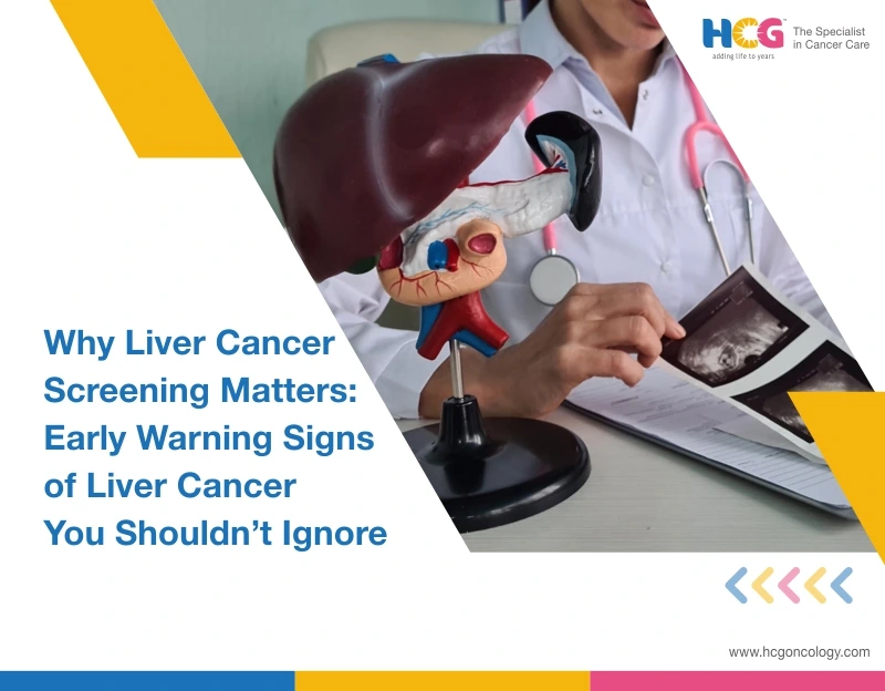
13 Apr, 2026
Feel free to reach out to us.
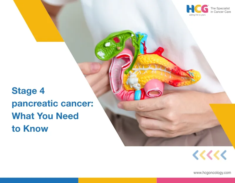
13 Apr, 2026
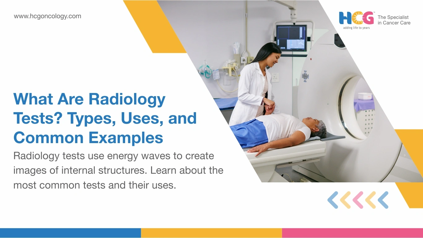
When your doctor says, "We need to run some imaging," what they mean is a radiology test. These are procedures that let the clinical team look inside your body at bones, organs, blood vessels, and soft tissue without making a single incision. The technology varies: some tests use X-rays, others rely on magnetic fields, sound waves, or a small amount of radioactive material. What they share is the ability to build a picture of your internal anatomy that no physical exam alone can provide.
A blood test tells your doctor what is happening chemically. A radiology test shows where something is happening and what it looks like. Both matter, but they answer different questions. And no two imaging tests are identical. Your doctor picks the right one based on which organ is involved, what condition is suspected, and how much detail the situation demands.
The below table provides estimate costs for different tests at hospitals like HCG Cancer Hospital:
| Test | Best Used For | Radiation? | Indian Cost Range |
|---|---|---|---|
| X-Ray | Fractures, chest infections | Low | ₹260 to ₹9,420 |
| CT Scan | Injuries, tumors, brain | Moderate | ₹6,747 to ₹32,590 |
| MRI Scan | Spine, soft tissue, joints | None | ₹8,120 to ₹32,960 |
| Ultrasound | Abdomen, pelvis, pregnancy | None | ₹1,600 to ₹14,800 |
| PET Scan | Cancer staging | Low (tracer) | ₹24,670 to ₹65,270 |
| Fluoroscopy | GI tract, stent placement | Real-time low | ₹4,500 to ₹11,000 |
Rates vary by hospital, city, and patient profile. Mumbai, Delhi, and Bangalore generally run higher than Tier 2 or Tier 3 locations.
Every imaging modality works on a different physical principle. That is why they are not interchangeable.
| Scan | Best Used For | Key Watch-Out |
|---|---|---|
| X-Ray | Fractures, chest infections, respiratory symptoms, quick bone assessment | Results same day; first scan ordered before anything advanced |
| Fluoroscopy | Tracing contrast through the bowel, guiding catheter or stent placement, live procedural navigation | Higher radiation than X-ray; used only when live imaging is clinically necessary |
| CT Scan (with contrast) | Trauma assessment, characterizing nodules, staging known cancer, cross-sectional organ detail | Flag iodine allergy or kidney concerns before contrast is given |
| MRI (with gadolinium) | Brain, spinal cord, soft tissue evaluation, tumor extent, neurological changes | Declare all implants before booking; pacemakers and cochlear devices may be incompatible |
| Ultrasound + Doppler | Abdominal organs, pelvic assessment, pregnancy monitoring, circulation checks | No radiation, no special preparation; Doppler added without extra steps when blood flow needs assessment |
| PET/CT | Metabolic staging before structural changes appear, treatment response, detecting recurrence | Maps activity, not anatomy alone; a biopsy still confirms cancer |
MRI and ultrasound involve no ionizing radiation. For most adults, the dose from an X-ray or CT is modest relative to the diagnostic value gained.
Pregnant patients, young children, and those with repeated imaging history require extra consideration. Always tell your radiology team if there is any chance of pregnancy before a scan involving radiation.
After a PET scan, basic radiation clearance steps apply for a few hours: keep a distance from young children and pregnant women while the radiotracer passes through urine. Post-scan hydration matters here. Drinking 1.5 to 2 liters of water in the hours following a PET scan or contrast-enhanced CT helps the kidneys clear the radiotracer or iodine dye faster. Your team will adjust this advice based on your kidney function.
If you experience MRI anxiety, please discuss it with us before your appointment. Most centers can offer a preparatory consultation or light sedation for claustrophobic patients, which indirectly improves image quality, since movement during the scan blurs results.
After your scan, a radiologist interprets the images and sends a written report to your referring doctor. How long this takes depends on the scan. A plain chest X-ray can be read in minutes. A full-body PET-CT or gadolinium-enhanced MRI typically takes 24 to 48 hours, given the reconstruction work and level of specialist review involved.
Ask your doctor to walk you through the report. Radiology language is written for clinicians, not patients, and misreading a raw report without context causes unnecessary worry. Request a digital copy of both the images and the written findings for your own records.
Each radiology test, whether it's contrast-enhanced CT, gadolinium MRI, Doppler imaging, or PET-CT with Time-of-Flight technology. It is built to answer a specific clinical question. The choice your doctor makes reflects what they need to see, how urgently they need to see it, and which technology gives the clearest answer for your situation.
HCG Cancer Hospital, a prominent cancer hospital in India, uses advanced diagnostic radiology across every cancer care pathway, including PET-CT with Time-of-Flight technology, MRI, Doppler imaging, and fully digital workflows. All imaging is reviewed by the HCG’s Radiology Team and integrated into multidisciplinary tumor board decisions.
Disclaimer: This information is intended to educate patients and caregivers. It does not replace professional medical advice. All treatment decisions should be made in consultation with a qualified doctor.