05 May, 2026
05 May, 2026
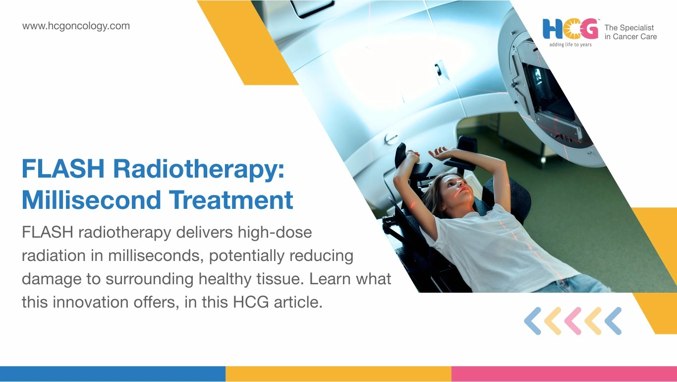
FLASH radiotherapy refers to Fast-acting, Low-toxicity, Accelerating, Safe, and High-dose-rate radiotherapy. It compresses a full therapeutic radiation dose into under one second, operating at dose rates exceeding 40 gray per second. Standard radiotherapy delivers that same dose over minutes per session across weeks of daily appointments. The speed difference is not cosmetic. Preclinical research consistently shows that this extreme acceleration appears to spare healthy tissue significantly while maintaining comparable tumor destruction. Human trials are underway, but widespread clinical availability has not yet arrived.
FLASH radiotherapy works by exploiting a biological asymmetry between cancer cells and healthy tissue. The result is the dose-rate effect, wherein tumor destruction proceeds at rates comparable to conventional radiotherapy, while surrounding organs experience measurably less collateral injury.
Speaking of benefits, such an approach could translate to fewer cases of fibrosis, mucositis, or late organ damage, the side effects that most concern patients completing long radiation courses.
| Method | Dose Rate | Delivery Time Per Session | Typical Session Count |
|---|---|---|---|
| Conventional radiotherapy | 0.03 to 0.05 Gy/second | 10 to 20 minutes | 20 to 35 sessions |
| Hypofractionated SBRT | 0.1 to 10 Gy/second | 3 to 10 minutes | 3 to 5 sessions |
| FLASH radiotherapy | Over 40 Gy/second | Under 1 second | Under investigation |
FLASH radiotherapy is not yet a routine clinical option. As of 2026, access is limited to early-phase trials at specialist international centers, with enrollment focused on specific sites, including bone metastases and select solid tumors. Broader availability depends on human trial outcomes followed by regulatory review.
The primary engineering barrier is modifying existing linear accelerators and proton systems to reach FLASH-range dose rates without losing spatial precision. Several manufacturers are developing FLASH-capable hardware. No platform has received widespread regulatory approval for routine clinical use at the time of writing.
Each platform undergoes extensive treatment planning, beam calibration, and dosimetry verification before any patient exposure. Spatial accuracy cannot be traded for speed.
Patients enrolled in FLASH trials follow structured aftercare protocols. Interval CT or MRI imaging at defined timepoints measures tumor response and screens for unexpected tissue reactions. For thoracic targets, pulmonary function testing is scheduled routinely.
Nutritional counseling and fatigue management address the symptoms that accompany any radiation-based regimen. Skin assessment monitors acute surface reactions at treated sites. Psychological support and caregiver resources are built into trial participation, given the additional uncertainty experimental treatment carries. A rehabilitation referral is available where a tumor site or treatment field affects physical function. All follow-up imaging schedules are set by the trial protocol and the treating radiation oncologist.
FLASH radiotherapy carries no standard commercial pricing in India at present, as patient access remains within trial structures only.
When FLASH platforms eventually reach commercial deployment, pricing will reflect equipment complexity, center specialization, and treatment protocol.
At HCG Cancer Hospital, radiation therapy starts with a straightforward premise: the right dose, delivered to precisely the right place, every session. Each patient's treatment plan is built from the ground up, factoring in tumor size, location, proximity to critical structures, and how the body responds at each stage of care.
Specialists work across a range of advanced radiotherapy platforms selected for specific clinical needs. CyberKnife enables frameless stereotactic radiosurgery, targeting tumors with sub-millimeter accuracy. Radixact allows clinicians to reshape treatment fields as tumor geometry changes over the course of therapy. And Ethos brings AI-driven adaptive planning into the mix, using imaging data from each session to generate a refined plan before the patient even leaves the treatment room.
That kind of flexibility (built into the workflow, not added as an afterthought) is what allows the team to respond to real-time anatomical changes rather than treating every session as identical to the first.
Disclaimer: This information is intended to educate patients and caregivers. It does not replace professional medical advice. All treatment decisions should be made in consultation with a qualified doctor.
Feel free to reach out to us.