10 Apr, 2026
Feel free to reach out to us.
10 Apr, 2026
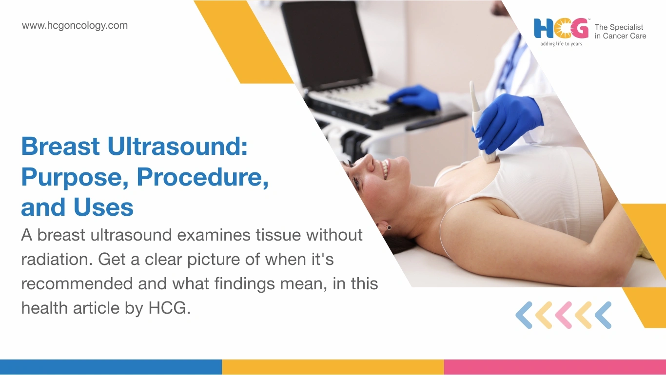
Breast ultrasound is a non-invasive imaging technique that involves high-frequency sound pulses to generate live, cross-sectional pictures of breast tissue, with absolutely no radiation involved. Physicians order this breast sonography test when a mammogram highlights a region of concern or when a patient reports noticing a lump. It is quick, painless, and preparation-free; the scan takes under 30 minutes. For women with dense breast tissue, where X-ray contrast fails to separate tumors from fibroglandular backgrounds, breast sonography often surfaces findings that change everything.
Sonographic breast examination works by directing rapid sound bursts through skin and glandular tissue via a probe called a "transducer," operating at 7.5 to 15 MHz for breast imaging. At every boundary between two tissue types, part of the signal bounces back. The machine translates those returning echoes into a moving, cross-sectional image on screen.
Denser material reflects more sound and registers brighter. Fluid absorbs the signal and appears uniformly dark. Radiologists precisely capture this contrast to map whatever sits beneath the surface.
Common Confusion: Mammography produces a flat radiographic shadow of the entire breast. Breast sonography allows radiologists to focus on one specific zone in three dimensions, rotating the probe to study a mass from multiple orientations without any radiation.
| Parameter | Breast Ultrasound | Mammography |
|---|---|---|
| Energy source | High-frequency sound (7.5–15 MHz) | Low-dose ionizing X-ray |
| Primary strength | Lump characterization, dense breasts | Microcalcifications, population screening |
| Dense breast sensitivity | Reliably strong | Meaningfully reduced |
| Suitable for | All ages | Typically recommended from age 40 |
| Biopsy guidance | Yes, real-time. | Limited |
| Scan duration | 15 to 30 minutes | 10 to 20 minutes |
In summary, mammography screens the whole breast broadly. Breast ultrasound examines with precision whatever clinical examination or prior imaging has already flagged.
Breast ultrasound is recommended when physical or radiographic findings need tissue-level clarification. Radiologists at HCG typically order breast ultrasounds when:
Good to Know: Most breast ultrasound scans end with a reassuring benign finding. The scan clarifies rather than automatically signaling something serious.
No dietary changes are recommended. No injections are involved. No compression whatsoever. Here is what actually happens:
Step 1: Positioning: You lie on a table with your arm raised on the relevant side. Breast tissue spreads naturally, improving transducer contact across the surface.
Step 2: Gel Application: A water-based acoustic coupling gel is applied to the skin. Air pockets between the probe and skin scatter sound signals; the gel eliminates them entirely, keeping images sharp.
Step 3: Real-time Sonography: The radiologist moves the transducer across the breast in deliberate, overlapping passes. The B-mode image refreshes continuously, allowing immediate redirection toward any suspicious zone.
Step 4: Reporting: Reporting dimensions, boundary characteristics, internal echo patterns, and posterior acoustic effects of any identified structure is formally done. Color Doppler assessment of vascular flow may be added when evaluating a solid nodule.
Good to Know: Eat, drink, and take all medications normally before the appointment. Absolutely nothing needs to change in your morning routine.
Distinguishing a benign fluid cyst from a solid nodule is arguably the single most clinically decisive function breast sonography performs.
| Feature | Fluid-Filled Cyst | Solid Nodule |
|---|---|---|
| Internal echoes | Absent (uniformly anechoic) | Visible throughout |
| Boundary characteristics | Smooth, sharply defined | Irregular or lobulated |
| Posterior acoustic effect | Enhancement behind the cyst | Shadowing in certain cases |
| Colour Doppler vascularity | Absent | May show internal blood flow |
| Typical clinical next step | Observation or aspiration | Biopsy if features are suspicious |
A simple cyst with clean margins, a uniform dark interior, and posterior acoustic enhancement carries a very low malignancy probability. A solid nodule showing spiculated edges and Doppler-detectable vascularity warrants an ultrasound-guided core needle biopsy for definitive histopathological assessment.
Know This: An abnormal scan does not confirm cancer. It identifies a structure that requires closer examination. Only pathological analysis of sampled tissue establishes a definitive diagnosis.
Breast ultrasound performs with high sensitivity when characterizing masses, particularly within dense fibroglandular tissue where mammographic contrast is insufficient. Clinical evidence reviewed by the American Cancer Society confirms that sonography detects breast cancers in dense-breast populations that standard X-ray screening overlooks.
Its honest limitation is that microcalcifications, tiny calcium deposits that can signal early ductal carcinoma in situ, frequently escape sonographic detection because sound-wave contrast does not reliably differentiate them. Operator skill and probe frequency selection also influence the diagnostic yield measurably.
HCG's breast imaging team pairs sonography with mammography or MRI for high-risk patients, applying a combined modality strategy to maximize sensitivity without unnecessary interventions.
Dense breast tissue holds more fibroglandular cells relative to fatty tissue. On a standard mammogram, dense glandular structures and many breast tumors register identically as white regions, creating an overlap that can effectively hide a lesion.
Breast sonography bypasses this problem entirely. Sound characterizes tissue based on acoustic properties, not X-ray absorption contrast. A tumor buried within a dense fibroglandular background becomes visible where the mammogram showed nothing of note.
A substantial proportion of Indian women have naturally dense breast composition. For this group, supplemental breast ultrasound is not a discretionary add-on. Clinically, it is often the step that reveals a finding that would otherwise remain undetected.
If a prior mammogram report described your breast tissue as heterogeneously or extremely dense, ask your physician directly whether adjunct sonography is appropriate for your situation.
The coupling gel wipes off in seconds. Patients resume all normal activities immediately, including work and exercise. A formal radiologist report reaches the referring physician within 24 to 48 hours.
Two pathways are typically followed:
Benign cyst confirmed: Scheduled follow-up imaging at 6 to 12 months, or aspiration if the cyst is causing discomfort. Post-procedure care involves minimal site hygiene only.
Solid or indeterminate nodule identified: Referral for ultrasound-guided core needle biopsy. After the biopsy, keep the site clean, avoid heavy lifting for 24 hours, and contact your care team promptly if unusual swelling or discharge develops.
HCG's patient navigators offer emotional support, second-opinion coordination, and multidisciplinary tumor board review for patients awaiting biopsy outcomes.
| City Tier | Approximate Cost Range |
|---|---|
| Metro cities (Mumbai, Delhi, Bengaluru) | ₹1,500 to ₹9,000 |
| Tier 2 cities (Vijayawada, Nagpur) | ₹800 to ₹3,000 |
Costs vary by hospital, its location, and patient profile. Always confirm the exact fee with your chosen facility before scheduling.
HCG Cancer Hospital places breast ultrasounds within a fully integrated imaging program, where radiologists work directly alongside oncologists and surgeons on every complex finding. Patients arrive carrying a question. They leave with a documented, clinician-reviewed answer and a clear pathway forward, supported at every step by HCG's comprehensive cancer care network.
Disclaimer:This information is intended to educate patients and caregivers. It does not replace professional medical advice. All treatment decisions should be made in consultation with a qualified doctor.