02 May, 2026
02 May, 2026
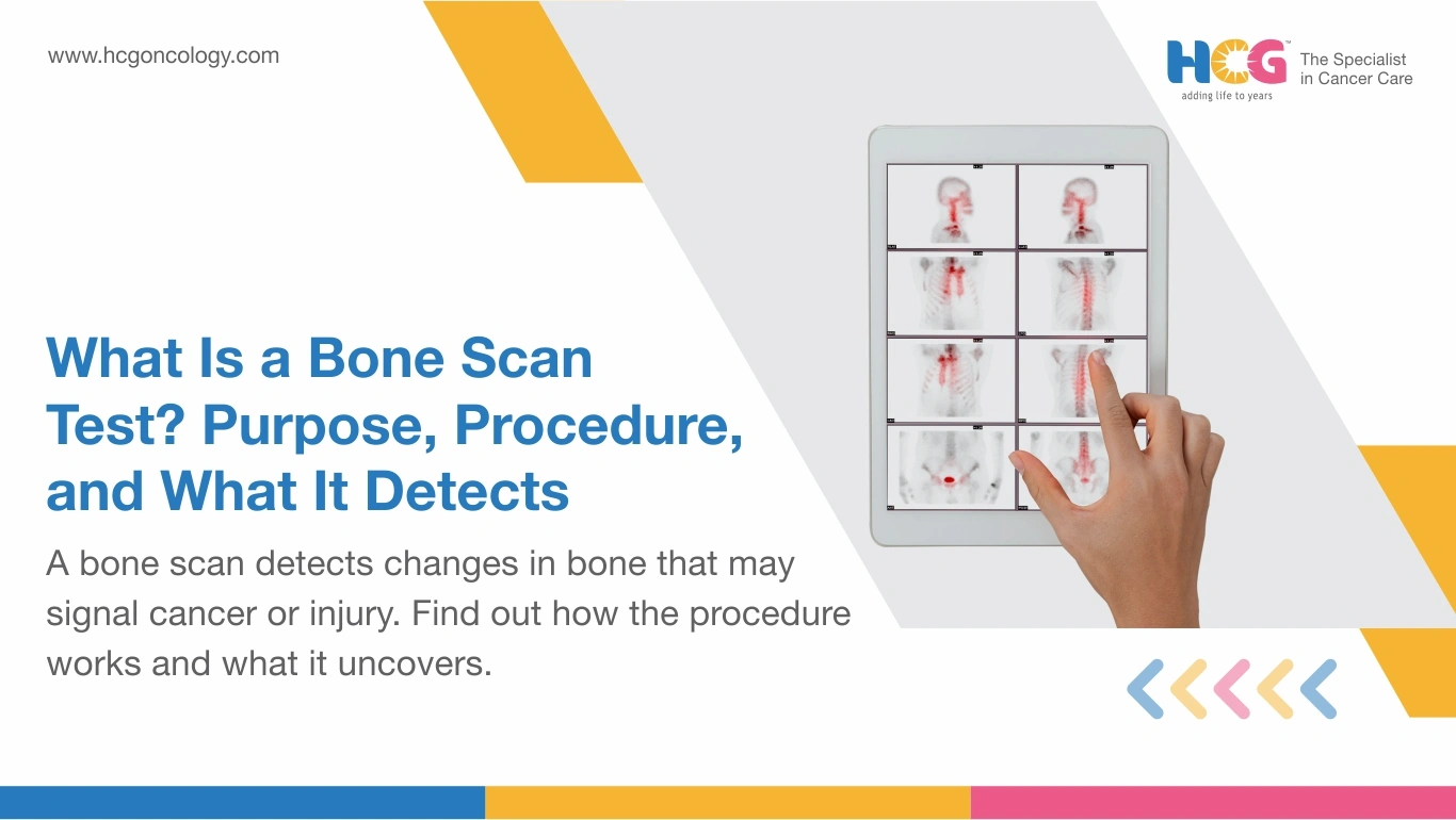
A bone scan test is a nuclear medicine imaging procedure that maps metabolic activity across the skeleton using a small radioactive tracer. Where X-rays capture what bones look like structurally, a bone scan reveals how bones are behaving biologically. Abnormal areas absorb the tracer differently, producing distinct signals on a gamma camera. Oncologists, orthopedic specialists, and infection medicine teams all rely on this test when standard imaging falls short.
A bone scan test maps how actively different regions of the skeleton are responding to biological stress, using a radioactive tracer to reveal what X-rays cannot.
Technetium-99m, the tracer used in most bone scans, travels through the bloodstream after intravenous injection and collects in areas of elevated bone activity. Tumors, fractures, infections, and inflammatory conditions all accelerate local bone metabolism. Those regions absorb more tracer and show up as brighter areas on the gamma camera image.
A bone scan can detect abnormalities several months before they become visible on a standard X-ray, which makes it a genuinely useful early-detection tool in the right clinical context.
A bone scan is ordered to detect cancer spread to bones, unexplained bone pain, stress fractures, osteomyelitis, inflammatory arthritis, and metabolic bone conditions such as Paget's disease.
| Condition | What the Scan Shows |
|---|---|
| Bone metastasis | Multiple hot spots across the skeleton |
| Stress fractures | Focal high tracer uptake at the fracture site |
| Osteomyelitis | Intense localised tracer concentration |
| Paget's disease | Irregular expanded areas of high activity |
| Arthritis | Symmetrical uptake patterns at joints |
| Avascular necrosis | Reduced or absent tracer uptake |
At HCG Cancer Hospital, nuclear medicine specialists interpret bone scan findings alongside blood markers and patient history before drawing any diagnostic conclusions.
A bone scan measures metabolic bone activity using a radioactive tracer. An MRI reveals structural soft tissue and bone detail using magnetic fields. An X-ray captures bone density changes only. Each serves a different diagnostic purpose.
| Feature | Bone Scan | X-ray | MRI |
|---|---|---|---|
| What it detects | Metabolic activity | Bone density changes | Structural soft tissue detail |
| Radiation | Low-dose tracer | Low-dose X-ray | None |
| Whole body coverage | Yes | No | No |
| Best for | Cancer spread, infection | Fractures, density loss | Soft tissue, marrow |
| Cost level (India) | Moderate to high | Low | High |
The bone scan test follows three stages:
Yes. The radiation dose is low, the tracer carries no allergy risk for most patients, and technetium-99m clears the body through urine within 24 to 48 hours.
Patients should drink extra fluids and urinate frequently in the hours after the scan to help flush the tracer. Breastfeeding mothers should pause nursing for a period as directed by their nuclear medicine physician. The procedure is generally avoided during pregnancy unless the clinical need is considered urgent and the risk-benefit assessment supports it.
No fasting is needed. Remove jewelry before the scan. Inform the team of recent fractures, surgeries, medications, or any chance of pregnancy before tracer injection.
Wear comfortable, loose clothing without metal fastenings. Patients who have recently undergone contrast imaging studies using barium or bismuth should inform the nuclear medicine team, as residual contrast can interfere with tracer uptake patterns on the scan image.
Activity resumes immediately after the scan. No recovery period is needed.
Drinking extra fluids for the first 24 hours speeds tracer elimination through urine. A follow-up appointment is typically scheduled within a few days for results review, though urgent findings are communicated sooner.
Bone scan results require specialist interpretation. A nuclear medicine physician reviews tracer distribution patterns and provides a written report, which the referring oncologist or orthopedic specialist then discusses with the patient. HCG's multidisciplinary imaging teams link scan findings directly to the patient's treatment pathway rather than issuing standalone reports.
| Condition | What the Scan Shows |
|---|---|
| Bone metastasis | Multiple hot spots across the skeleton |
| Stress fractures | Focal high tracer uptake at the fracture site |
| Osteomyelitis | Intense localized tracer concentration |
| Paget's disease | Irregular expanded areas of high activity |
| Arthritis | Symmetrical uptake patterns at joints |
| Avascular necrosis | Reduced or absent tracer uptake |
At hospitals like HCG Cancer Hospital, bone scan tests may cost from ₹5,950 to ₹25,130, depending on the type of scan (standard, 3-phase, or SPECT-CT), scope of imaging, and the city and center where it is performed.
Government facilities may offer bone scans at subsidized rates. Pricing in metropolitan cities like Mumbai, Delhi, Bangalore, and Chennai differs from Tier-2 and Tier-3 locations. Lastly, it is essential to note that costs vary by hospital and patient profile.
A bone scan test delivers a level of whole-body diagnostic detail that neither X-rays nor blood tests can replicate, particularly for detecting bone metastasis, infection, and metabolic bone disease at an early stage. The procedure is safe and completed within a single outpatient visit and well-tolerated by most adults. For patients managing a cancer diagnosis, unexplained bone pain, or an unresolved orthopedic concern, discussing whether a bone scan is the right next step with a nuclear medicine specialist is a practical, low-risk decision that can meaningfully sharpen the clinical picture.
Disclaimer: This information is intended to educate patients and caregivers. It does not replace professional medical advice. All treatment decisions should be made in consultation with a qualified doctor.
Feel free to reach out to us.