A PET-CT scan is a single imaging test that combines PET (positron emission tomography) with CT (computed tomography) to support cancer diagnosis, staging, and treatment planning.
At HCG centers in India, this combined scan, often shortened to "PET scan," shows how actively cells are using energy, while CT shows the exact size, shape, and location of that activity.
A PET scan alone captures only the metabolic picture; combining it with CT is what lets a specialist match that activity to a specific organ or structure. On its own, a PET-CT scan cannot confirm cancer.
Unusual activity still needs to be read alongside other findings, and sometimes a biopsy, before a diagnosis is made.
HCG PET-CT Scan centers in India
HCG provides PET-CT scanning at selected centers in India. Explore the verified HCG PET-CT scan centers below to confirm local availability, PET-CT technology, scan protocols, and appointment requirements. Services may differ by hospital, so patients should confirm the prescribed scan with the preferred center before booking.
What Is a PET-CT Scan?
A PET-CT scan is a single imaging examination that produces two different kinds of information at the same time. PET stands for positron emission tomography. CT stands for computed tomography.
The PET component is a form of functional imaging. It shows how actively cells in a particular area are using energy. Before the scan, a small amount of a radiotracer is given, usually into a vein. A radiotracer is a substance the body uses naturally, such as a form of sugar, tagged with a low dose of a radioactive material. Cells with higher metabolic activity take up more of the tracer, and the PET scanner records where that activity is concentrated.
The CT component is anatomical imaging. It uses X-rays to build detailed cross-sectional pictures of organs, tissues, and bones, which show size, shape, and exact position.
Software then aligns the two sets of images. The result is a single view in which an area of altered metabolic activity can be matched to a specific anatomical location.
This matters in cancer care because activity alone is not enough to act on. Knowing that a region is metabolically active is useful. Knowing exactly which lymph node, which segment of the liver, or which part of the lung it corresponds to is what allows a specialist to plan a biopsy, a surgery, or a radiation field.
Is a PET Scan the Same as a PET-CT Scan?
Not exactly, although the terms are often used interchangeably in everyday conversation.
A PET scan on its own shows metabolic or functional activity. A PET-CT scan combines those PET findings with CT-based anatomical detail in one session, on one machine.
In practice, most PET imaging performed for cancer today is PET-CT, which is why many patients and even some referral notes say "PET scan" when the examination being arranged is a combined PET-CT.
Patients should confirm the exact test prescribed with the treating specialist or the scan center when booking, as preparation and reporting can differ.
Why Is a PET-CT Scan Done?
A PET-CT scan may be recommended at several points in cancer care. It is ordered by a treating specialist for a defined clinical reason, not as a routine test.
Common reasons include:
- Investigating a suspicious finding seen on another scan or test
- Supporting the diagnosis of certain cancers, alongside other investigations
- Staging a confirmed cancer, which means assessing how far it has progressed
- Checking whether cancer has spread to lymph nodes or distant organs
- Helping plan the most appropriate treatment approach
- Supporting radiation therapy planning by defining the area of active disease
- Assessing how well a cancer has responded during or after treatment
- Investigating a possible recurrence when symptoms or test results raise concern
- Identifying the most informative site for a biopsy in selected cases
PET-CT supports these decisions. It does not replace them. A specialist reads the scan together with the clinical history, physical examination, laboratory results, and, where relevant, pathology findings.
What Can a PET-CT Scan Detect?
A PET-CT scan can show:
- Areas of unusually high or unusually low metabolic activity
- Certain primary cancers, depending on type and location
- Whether disease has spread beyond the original site
- Disease that remains after treatment, or disease that has returned
- Metabolic changes that may reflect how a tumor is responding to treatment
- Selected non-cancer conditions, such as some infections and inflammatory disorders, where clinically relevant
An important qualification: increased tracer uptake does not always mean cancer. Infection, inflammation, and normal organ activity may also cause increased uptake, so PET-CT findings must be interpreted by a qualified specialist alongside the patient's history and other test results.
The reverse is also true. Some cancers are slow-growing or have low metabolic activity, and these may show little uptake even when disease is present.
Types of PET-CT Imaging
The radiotracer and PET-CT protocol selected depend on the cancer type and clinical indication. HCG centers may offer different PET-CT technologies and scan protocols depending on local availability.
FDG PET-CT
FDG stands for fluorodeoxyglucose, a form of glucose tagged with a radioactive marker. Because many cancer cells consume glucose at a higher rate than surrounding tissue, FDG tends to concentrate in metabolically active areas.
FDG PET-CT is the most widely used form of PET imaging in oncology and is applied across a range of cancer types for staging, response assessment, and recurrence evaluation. Its usefulness varies by tumor type.
PSMA PET-CT
PSMA stands for prostate-specific membrane antigen, a protein found in higher concentrations on many prostate cancer cells.
PSMA PET-CT is used in selected prostate cancer situations, such as assessing the extent of disease or investigating a rising PSA level after treatment. Whether it is appropriate depends on the clinical stage and the treating specialist's assessment.
DOTATATE or DOTANOC PET-CT
These tracers bind to somatostatin receptors, which are present in higher numbers on many neuroendocrine tumor cells.
This form of imaging is used in selected neuroendocrine tumor cases to locate the primary tumor, assess spread, and help determine whether certain receptor-targeted treatments may be suitable.
Digital PET-CT
Digital PET-CT uses newer detector technology that can improve image clarity and may allow imaging at a lower tracer dose in appropriate cases.
Availability differs by center. You can read more about this on our dedicated page covering Digital PET-CT technology.
How Does a PET-CT Scan Work?
The examination follows a predictable sequence:
- A small amount of radiotracer is administered, most often through a vein in the arm.
- The patient rests quietly while the tracer distributes through the body.
- Areas with different metabolic activity absorb different amounts of the tracer.
- The PET scanner records where tracer activity is concentrated.
- The CT scan captures the anatomical detail of the same region.
- The two image sets are combined and interpreted by a nuclear medicine specialist.
The waiting period after the tracer is given is often referred to as the uptake phase. Patients are usually asked to stay still and avoid talking or moving unnecessarily during this time, because muscle activity can draw tracer into the muscles and affect image quality.
Before You Book a PET-CT Scan
Have the following ready when you contact your preferred HCG center:
- The doctor's prescription or referral, stating the clinical indication
- Previous imaging reports and scans, for comparison
- A full list of current medicines
- Whether you have diabetes, so appointment timing can be planned around it
- Whether you are pregnant, think you might be, or are breastfeeding
- Your preferred city or HCG center
The center uses this to confirm the correct scan type and radiotracer, check technology availability, and plan the uptake-period timing before your visit.
How to Prepare for a PET-CT Scan
Preparation instructions come from the center performing the scan and should be followed closely, since image quality depends on them.
Points typically covered include:
- Fasting. Patients are usually asked not to eat for several hours before the scan, since food affects blood sugar and can alter tracer uptake.
- Water.Plain water is generally allowed and often encouraged. Sweetened drinks are not.
- Exercise. Strenuous activity in the day before the scan is usually discouraged, as it increases muscle uptake.
- Current medicines. Bring a full list. Do not stop any medicine unless specifically instructed.
- Diabetes. Blood glucose levels influence FDG PET-CT quality. Patients with diabetes should tell the center in advance so that timing, medication, and insulin can be planned.
- Pregnancy. Tell the team if you are pregnant or think you might be.
- Breastfeeding. Tell the team, as specific guidance on feeding after the scan may be needed.
- Allergies. Report any previous reaction to contrast media or other injections.
- Kidney function. If contrast is planned, a recent kidney function test may be requested.
- Previous scans and records. Bring earlier imaging, reports, and the referring prescription. Comparison with prior scans is often central to interpretation.
- Clothing. Wear comfortable clothing without metal fasteners. You may be asked to change into a gown.
- Arrival time. Arrive at the time given, which is usually earlier than the scan itself to allow for registration and the uptake period.
Preparation can vary depending on the radiotracer, scan protocol, and medical condition. Patients should follow the instructions provided by the PET-CT center.
What Happens During and After a PET-CT Scan?
Before the scan
Registration is completed, and a member of the team reviews the medical history, current medicines, and the referring prescription. Where required, blood glucose is checked. The radiotracer is then administered, usually through a vein. The patient rests quietly during the uptake period.
During the scan
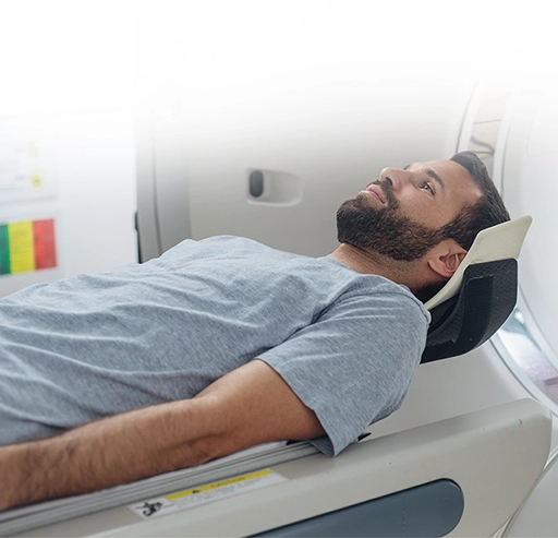
The patient lies on a scanning table that moves slowly through the ring-shaped scanner. Remaining still is important, since movement blurs the images. PET and CT images are acquired in the same session. In some cases, CT contrast is given when it is clinically required. The scanner is open at both ends and does not touch the patient.
The team stays in contact throughout, usually through an intercom, and can be reached at any point.
After the scan
Most patients resume routine activities unless they are told otherwise. Drinking fluids is usually advised, as this helps the body clear the remaining tracer. Some centers advise limiting close, prolonged contact with infants and pregnant women for a few hours as a precaution. The images are then reviewed and reported by a specialist, and the findings are discussed at the follow-up consultation.
How Long Does a PET-CT Scan Take?
Patients often ask about the scan time and are surprised that the appointment is longer than the imaging itself. It helps to think of the visit in four parts:
- Registration and preparation, including history review and any blood glucose check
- The radiotracer uptake period, during which the patient rests quietly
- The actual scanning time, which is usually the shortest part of the visit
- Possible additional or delayed imaging, if the specialist requests further views
Total time varies by protocol, tracer, and individual clinical requirements, so centers generally give an expected duration at the time of booking rather than a fixed figure.
Is a PET-CT Scan Safe?
PET-CT is generally considered safe when clinically indicated and performed using appropriate protocols.
The examination does involve ionizing radiation from both the radiotracer and the CT component. The tracer dose is controlled and kept to the level needed for a diagnostic-quality image. Before a scan is recommended, the expected clinical benefit is weighed against that exposure.
A few situations need specific discussion:
- Pregnancy. PET-CT is generally avoided during pregnancy unless there is a compelling clinical reason. Tell the team before the scan.
- Breastfeeding. Specific guidance on timing may be given.
- Contrast. If CT contrast is planned, previous reactions and kidney function are reviewed first.
- Allergies. Any known allergy should be reported at registration.
Reactions to the radiotracer itself are uncommon. The injection feels much like a routine blood test, and the scan is not painful.
After the scan, patients may be given simple precautions to follow for a short period, such as drinking fluids and limiting extended close contact with young children.
What Do PET-CT Scan Results Mean?
A PET-CT report describes where tracer uptake is higher or lower than expected and what the CT images show at those locations.
Reports often mention SUV, or standardized uptake value. This is a number that expresses how much tracer has concentrated in a particular area relative to the rest of the body. It is one input among several. A higher SUV can indicate more active tissue, but it does not by itself confirm cancer, and values are not directly comparable across different scanners or protocols.
Comparison with previous imaging is often the most useful part of the interpretation, because a change over time can be more meaningful than a single measurement.
Scans are reviewed by a nuclear medicine specialist and read alongside the clinical history, examination findings, laboratory results and, where available, biopsy findings. At HCG, imaging is reviewed as part of the wider cancer-care pathway rather than in isolation.
Reporting time varies by center and by whether additional imaging or comparison studies are needed. Patients should ask the center when the report will be available and should discuss the findings with their treating specialist rather than interpreting numbers independently.
PET-CT vs PET Scan, CT Scan, and MRI
| Imaging test | Primarily shows | Common role |
|---|---|---|
| PET scan | Metabolic or functional activity | Shows areas with altered tracer uptake |
| CT scan | Anatomical structure | Shows the size and location of abnormalities |
| PET-CT | Metabolic activity plus structure | Combines PET findings with their anatomical location |
| MRI | Detailed soft-tissue anatomy | Useful for selected organs and clinical situations |
These tests are complementary rather than competing. No single modality is better than the others in every situation. The right choice depends on the cancer type, the organ being assessed, the clinical question being asked, and what previous imaging has already shown. Many patients undergo more than one type of scan over the course of their care.
Benefits and Limitations of PET-CT
Benefits
- Combines functional and anatomical information in one examination
- Supports accurate cancer staging
- May identify disease in more than one area of the body during a single session
- Assists treatment planning, including radiation therapy planning
- Helps assess how a cancer is responding to treatment
- May reduce the need for certain additional investigations
Limitations
- Not every cancer shows significant tracer uptake
- Infection and inflammation may also produce increased uptake
- Very small abnormalities may not be clearly visible
- Blood glucose levels can affect the quality of certain PET-CT scans
- Findings must be interpreted together with other clinical evidence
- PET-CT is not a routine population-screening test for every cancer
Understanding both sides helps set realistic expectations before the scan and makes the follow-up discussion more useful.
PET-CT Scan Cost
PET-CT scan charges can vary depending on the radiotracer, scan protocol, use of contrast, clinical indication, and HCG center. Patients should contact their preferred center with the prescription to obtain a current estimate and confirm what is included.
Why Choose HCG for a PET-CT Scan?
PET-CT at HCG is delivered inside a dedicated cancer-care network, not as a stand-alone scanning service.
Nuclear medicine expertise. Scans are acquired and reported by nuclear medicine specialists working within HCG's oncology centers, not a general radiology pool.
PET-CT equipment. The network operates 21 PET-CT scanners, including an in-house cyclotron at HCG Cancer Hospital, K. R. Road, Bengaluru, supporting on-site production of certain radiotracers (figure from the PET-CT cost page confirm still current before publishing).
Oncology integration. Findings are read alongside the patient's existing diagnosis, prior imaging, and pathology rather than as an isolated test.
Multidisciplinary review. Complex or borderline findings can go to HCG's multidisciplinary tumor board for joint review by medical, surgical, and radiation oncology.
Report delivery. Reports are issued by the reporting center and shared with the treating specialist for discussion at the follow-up consultation.
Center-specific services. Scan types and radiotracers differ by location; check the confirmed technology at your preferred center above before booking.