02 May, 2026
02 May, 2026
Bone cancer develops when skeletal cells lose their normal growth regulation and begin dividing without stopping. Two broad categories exist. Primary bone cancer starts inside the bone tissue itself. Secondary bone cancer occurs when a tumor from the breast, lung, prostate, or kidney sends malignant cells through the bloodstream into the skeleton.
Primary bone cancer is rare, under 1% of all cancers globally. Yet it carries serious weight, partly because it strikes children and young adults more than most malignancies. Knowing what to look for and when to act on it genuinely shapes how treatment goes.
Bone cancer takes hold when osteoblasts, chondrocytes, or primitive mesenchymal cells begin multiplying in a disorganized, unchecked way, eroding normal skeletal architecture from within.
Common confusion: Finding a tumor in a bone does not automatically mean bone cancer. Most bone tumors are benign and localized and carry no metastatic risk.
One distinction matters enormously here. A tumor that began inside the bone is primary. A tumor that migrated from the breast, kidney, or prostate is a bone metastasis, a secondary condition with a completely different treatment logic. The two are not interchangeable diagnoses, and conflating them leads to misaligned expectations about care.
| Type | Cell Origin | Typical Age | Common Sites |
|---|---|---|---|
| Osteosarcoma | Osteoblast cells | 10–30 years | Knee, upper arm |
| Ewing Sarcoma | Primitive mesenchymal cells | 5–20 years | Pelvis, long bone shafts |
| Chondrosarcoma | Chondrocyte cells | 40–70 years | Pelvis, shoulder, upper femur |
Osteosarcoma is the most common primary bone malignancy. It usually forms near growth plates, particularly around the knee. Chemotherapy before surgery is standard practice: it reduces tumor volume and, critically, reveals how responsive that specific tumor is to drug treatment. That response score guides every decision that follows.
Ewing sarcoma carries a defining chromosomal change called the EWS-FLI1 translocation. It responds well to both chemotherapy and radiation, which gives oncologists genuine flexibility in designing treatment, especially for younger patients, where preserving function matters greatly.
Chondrosarcoma follows its own rules. Standard chemotherapy barely affects it. Surgical removal with wide, clean margins is the primary strategy. Any tumor tissue left behind substantially raises the recurrence risk.
Good to Know: When cancer spreads to bone from another organ, treatment focuses on the original cancer's biology, not on bone-specific protocols.
No single confirmed cause exists. Several risk factors appear consistently across published research.
Inherited genetic conditions carry real weight. The RB1 mutation linked to hereditary retinoblastoma significantly raises osteosarcoma risk. Li-Fraumeni syndrome, rooted in TP53 mutations, broadens susceptibility across several cancer types, bone sarcomas among them.
Prior radiation to bone, especially in childhood, can trigger a secondary malignant tumor in the irradiated field years or even decades afterward.
Rapid adolescent growth correlates with peak osteosarcoma incidence. Fast-dividing osteoblasts during growth spurts appear to create windows of cellular vulnerability.
Certain pre-existing bone conditions, including Paget's disease and hereditary multiple osteochondromas, carry a measurable, though still small, risk of malignant change over time.
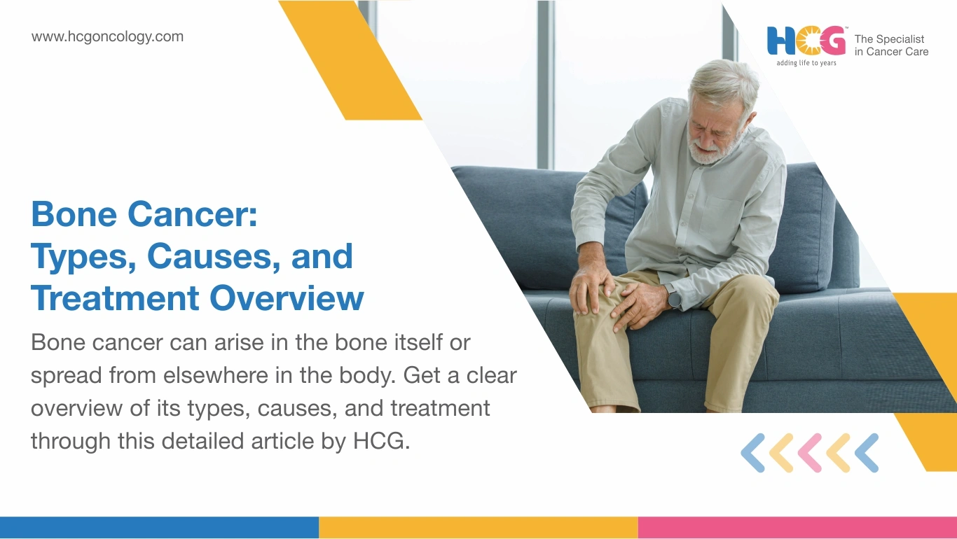
Bone cancer pain has a texture that distinguishes it from athletic injuries or wear-and-tear soreness. Patients describe it as a deep, dull, grinding ache that steadily intensifies and is characteristically worse at night, unresponsive to positional change or over-the-counter pain relief.
Beyond pain, pay attention to:
In summary, bone pain with no clear traumatic cause that persists beyond two weeks warrants imaging and a specialist review, not continued monitoring at home.
No single investigation confirms bone cancer alone. Diagnosis builds through a sequence.
Treatment is always individualized. Tumor subtype, grade, location, stage, and patient age all shape the plan. Decisions go through a multidisciplinary tumor board before any intervention begins.
Limb-salvage surgery has overtaken amputation as the standard for most extremity tumors. The surgeon removes the tumor with clear margins and rebuilds the limb using a custom prosthesis, bone graft, or both, preserving function alongside oncological control.
Chemotherapy anchors osteosarcoma and Ewing sarcoma treatment. Neoadjuvant chemotherapy before surgery shrinks the tumor and tests drug sensitivity simultaneously. A strong histological response is among the most favorable prognostic signals a clinician can see.
Radiation therapy is most relevant for Ewing sarcoma. It serves as a definitive local treatment when surgery is not feasible or as adjuvant therapy when margins are close after resection.
Personalized treatments meet the specific health needs of the patient. For instance, chondrosarcoma relies almost entirely on surgery. Wide margins are non-negotiable. Targeted agents such as sorafenib and regorafenib show some activity in refractory osteosarcoma through clinical trial access.
Rehabilitation after limb-salvage surgery starts within days. Physiotherapy around a new prosthetic joint takes months of consistent effort. Progress is real, but it is not fast.
Prostheses need scheduled radiographic checks, particularly in younger patients returning to high activity levels. Nutritional support through chemotherapy addresses the gap between dropping appetite and rising protein demand. An oncology dietician adjusts this through each phase.
Emotional recovery deserves the same attention as physical rehabilitation. Bone cancer tends to arrive when patients are building their lives. Psycho-oncology support, peer connections, and family counseling belong inside the care plan, not outside it.
Surveillance imaging, typically every three to six months after treatment ends, monitors for recurrence while intervention options remain viable.
| Treatment Component | Estimated Range (INR) |
|---|---|
| Diagnostic workup (MRI, biopsy, PET-CT) | ₹40,000 – ₹1,20,000 |
| Limb-salvage surgery with prosthesis | ₹5,00,000 – ₹15,00,000 |
| Chemotherapy per cycle | ₹30,000 – ₹1,50,000 |
| Radiation therapy full course | ₹2,00,000 – ₹6,00,000 |
Costs at hospitals in metro cities such as Bangalore, Mumbai, and Delhi generally sit at the upper range. Patients eligible under Ayushman Bharat PM-JAY may access partial coverage for select procedures. Costs vary by hospital and patient profile.
Bone pain lasting beyond two weeks without a clear injury explanation calls for action, not waiting.
In cancer care, HCG focuses on the reality that osteosarcoma, chondrosarcoma, and Ewing sarcoma are three distinct diseases requiring three distinct strategies. Placing each patient through a dedicated multidisciplinary tumor board, combining molecular diagnostics, ortho-oncology surgical expertise, and precision radiation delivery, is how individualized care actually gets built rather than simply promised.
Early specialist evaluation remains the variable that matters most.
Disclaimer: This information is intended to educate patients and caregivers. It does not replace professional medical advice. All treatment decisions should be made in consultation with a qualified doctor.
Feel free to reach out to us.