25 Aug, 2026
25 Aug, 2026
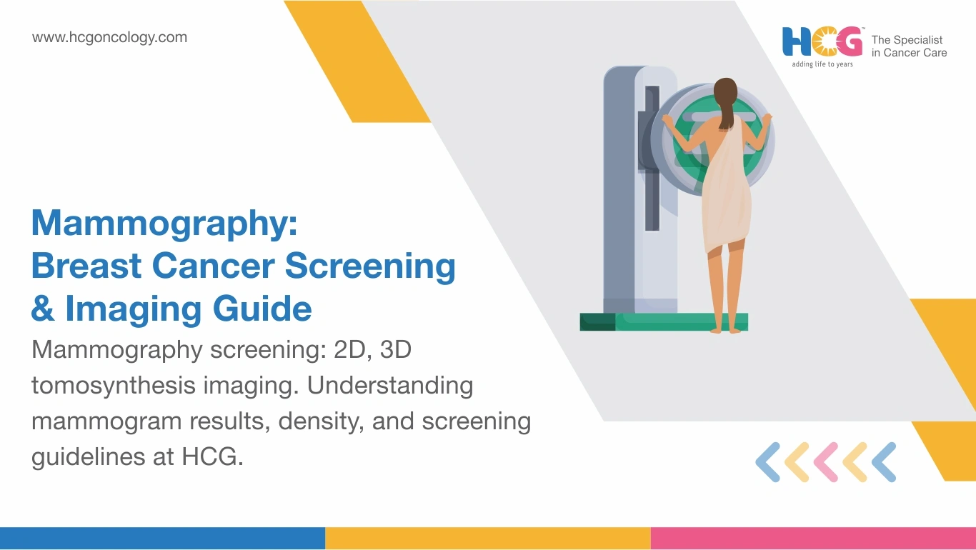
A mammogram is an examination used in screening for breast cancer by utilizing low levels of ionizing radiation through the breast to spot changes that would have gone unnoticed by other methods. They help locate lumps in the breasts that cannot be felt.
This is a very efficient means of screening for breast cancer despite the minor discomfort that comes with the process. Studies show that regular mammography screening can reduce breast cancer mortality by up to 20–40% in women aged 40 and above.
Mammography is a breast imaging technique designed to detect abnormal tissue changes before they can be felt or cause symptoms. It is the most effective screening tool for early-stage breast cancer detection in women over 40.
Your breast is placed on a flat surface and gently compressed between two plates.
Why the compression? It spreads the breast tissue so the image comes out clearer and the radiation dose stays low.
How long does mammography take? About 20 minutes total, though each compression lasts only a few seconds.
Is mammography painful? Most women describe pressure rather than sharp pain, and the feeling passes quickly. Scheduling your mammogram during the week after your period can cut down on tenderness because breast tissue is less sensitive at that point in the cycle.
Can mammography detect cancer early? Yes. Breast radiology teams use mammography to find breast calcifications (calcium deposits), microcalcifications (tiny calcium specks) and small masses that a physical exam would miss entirely. Using a magnifying glass to read fine print. Your hands couldn't feel what the image reveals.
Two main categories exist, and the difference matters.
But technology differences between mammography systems add another layer. Digital mammography captures images electronically instead of on film, letting radiologists adjust contrast for better detail. 3D mammography, also called tomosynthesis, takes multiple images from different angles to build a layered view of breast tissue.
Dense breast tissue makes it harder to read mammograms since both dense tissue and some breast abnormalities show up as white on the images.
For women with dense breasts, options like digital breast tomosynthesis, also known as 3D mammography, as well as breast ultrasound or MRI, might help. These extra screenings depend on each person’s risks. Always ask your healthcare provider for the best screening plan based on your specific situation.
Different tools are built for different purposes - that's the simplest way to put it. A mammogram uses X-rays to capture images of the entire breast. An ultrasound uses sound waves to examine specific areas in greater detail.
Mammography vs. MRI for breast cancer detection is another comparison that comes up often. MRI provides the most detailed imaging but isn't used for routine screening because it's expensive and produces more false positive mammogram results.
Mammography during pregnancy is generally avoided unless clinical concern requires it. The radiation dose is low, but doctors prefer ultrasound as the initial imaging tool during pregnancy because it doesn't involve radiation. If a breast lesion needs evaluation, your breast oncology team weighs risks and benefits carefully. Pregnancy doesn't prevent a mammogram from happening. It just shifts the decision-making process.
Age 40 is the starting line for regular mammography screening. How often should mammography be done? Current guidelines recommend every one to two years, depending on risk factors. Women carrying a family history of breast cancer or known genetic mutations may need to start earlier than 40.
Mammography is highly accurate for detecting early-stage breast cancer, though accuracy varies depending on breast tissue density; women with denser tissue may have a lower detection rate. A mammogram for breast cancer detection remains the gold standard in screening.
It can, particularly in women with dense breast tissue where tumors blend into the surrounding tissue on the image. That's why supplemental screening with ultrasound or MRI is sometimes recommended for high-risk patients.
And while mammography isn't perfect, it's the best routine screening tool available.
The BI-RADS score (Breast Imaging Reporting and Data System) is the standardized system used for reporting results, scaled from 0 to 6. A score of 1 means normal. A score of 0 means the images need further evaluation. Scores of 4 or 5 point to findings suspicious enough for a breast biopsy.
The follow-up process has structure.
At this stage, your case may be referred to a breast oncology team for further evaluation and a treatment plan if cancer is confirmed. A false positive mammogram creates stress, but it's common. Many abnormal findings turn out to be benign (non-cancerous).
The oncologists and radiologists working in the HCG Cancer Hospital conduct mammogram screenings through digital and 3D imaging methods that incorporate all screening and diagnosis procedures in one site. Patients can access all services provided in one location without having to make repeat appointments for follow-ups and biopsies.
The use of advanced techniques such as tomosynthesis helps detect abnormalities in dense breasts. Mammography screening is made easier through the scheduling program that the hospital has in place. Book a mammography screening appointment at your nearest HCG Cancer Hospital center.
Disclaimer: This article is for general informational purposes only and is not a substitute for professional medical advice, diagnosis, or treatment. Please consult a qualified healthcare provider for any questions regarding a medical condition.
Feel free to reach out to us.